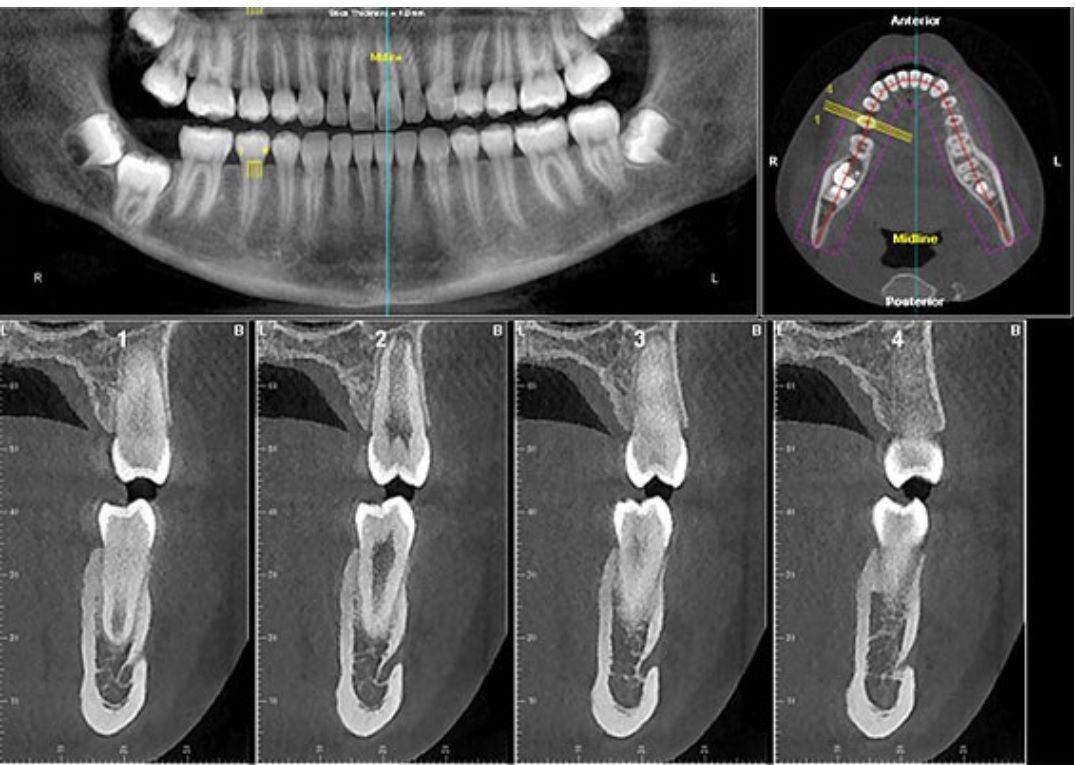

Intraoral techniques enable clinicians to clearly see the dentition and supporting structures, as well as diagnose alveolar bone loss and dental caries. Dental radiographs include intraoral techniques, such as bitewing and periapical images, along with extraoral techniques such as the lateral cephalometric and panoramic images. Radiographs allow clinicians to see conditions invisible to the naked eye and, when combined with oral assessments, provide valuable information about patients’ health.

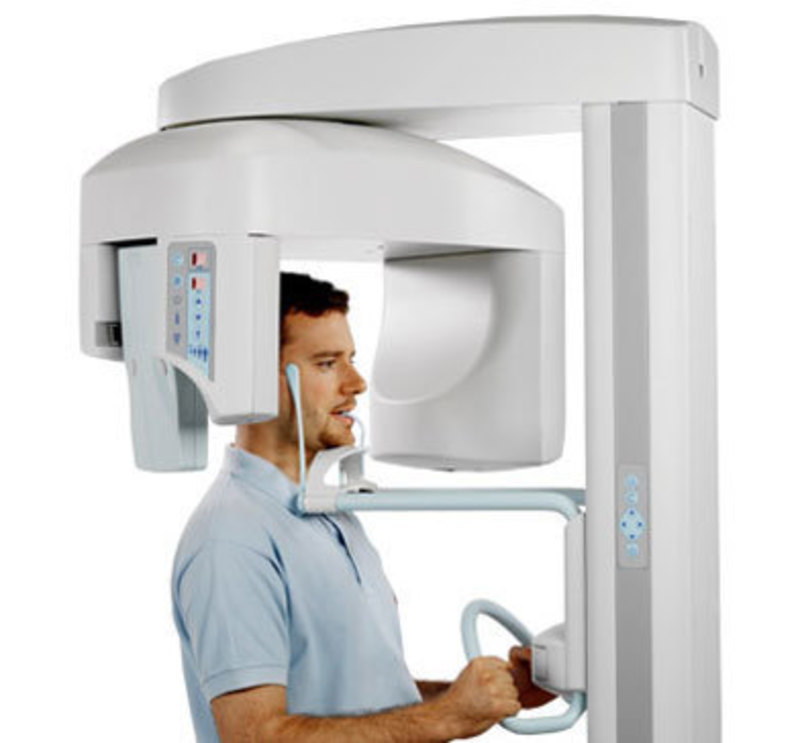
An OPG is just one diagnostic tool your dentist will use to ensure you maintain your dental health and prevent unnecessary dental issues.This course was published in the June 2020 issue and expires June 2023. This can be as part of a dental check up, or seperate appointment. The image is produced within seconds of the OPG being taken and your dentist will review it with you at the end of the appointment. Where can I get an OPG taken?ĭarlinghurst Dental has an OPG machine on site for your convenience and the images are produced digitally and stored in your digital file with all your other x-rays. The OPG usually takes about 3 mins to complete, only 20 seconds of this are the image being taken.

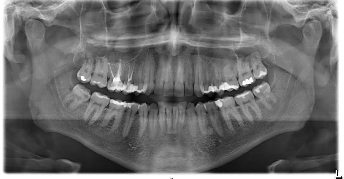
Your dentist will ask you to remove any jewellery or metallic items from around the head and neck area as it can interfere with the image, blocking the x-ray beam. Useful visual aid in patient education and case presentation.Short time required for producing the image.Ability to be used in patients who are restricted in opening their mouth.Convenience of examination for the patient.Broad coverage of facial bones and teeth including the TMJ (temperomandibular joint).The principal advantage of panoramic images/OPGs: It is also often used to check your jaw joint, the TMJ (temperomandibular joint), sometimes called the CMA (cranio-mandibular articulation), especially if you grind your teeth. This can be particularly useful to check hard to see areas like wisdom teeth, or the development of a child’s jaw and teeth, useful for assessing for development generally, but also orthodontic need. It shows less fine detail, but a much broader area of view. It is different from the small close up x-rays dentists take of individual teeth. At some stage in your dental treatment, your dentist will likely take an OPG.Īn OPG also demonstrates the number, position and growth of all the teeth including those that have not yet surfaced or erupted through the gum. The structures that are outside the viewed area are blurred. Panoramic x-rays allow images of multiple angles to be taken to make up the composite panoramic image, where the maxilla (upper jaw) and mandible (lower jaw) are in the viewed area. It shows a flattened two-dimensional view of a half-circle from ear to ear.

It is also sometimes called Orthopantomagraph or by the proprietary name Panorex. An OPG (Orthopantomagram) is a panoramic scanning dental X-ray of the upper and lower jaw.


 0 kommentar(er)
